Rib Cage Muscles Diagram - Muscles Of The Thoracic Wall 3d Models Video Tutorials Notes Anatomyzone / See more ideas about anatomy, anatomy study, rib cage anatomy.
byAdmin•
0
Rib Cage Muscles Diagram - Muscles Of The Thoracic Wall 3d Models Video Tutorials Notes Anatomyzone / See more ideas about anatomy, anatomy study, rib cage anatomy.. These rib muscles automatically get worked when you do bench presses, push ups and dips, but a few bonus exercises can help you really zero in for a more chiseled torso. On the dorsal side there is a neural spine. Further, there are two superior and two inferior processes meant for articulation with the neighbouring vertebra. These bony projections are used for attachment of muscles. Feel free to search our website for more information on this particular topic.
If you were to develop well defined rib cage muscles, they would give off the appearance of fingers on your sides. The fibres pass superolaterally to insert into the costal cartilages of muscles of the spine and 8 rib muscles anatomy rib muscles anatomy and human anatomy muscles rib cage diagram. Ribs, respiratory muscles and respiratory system | researchgate, the professional network for scientists. This post is about rib cage. Feel free to search our website for more information on this particular topic.
8 Muscles Of The Spine And Rib Cage Musculoskeletal Key from musculoskeletalkey.com Great diagram showing the positions of the deltoid and the tricep from the back. Each articulates with a thoracic vertebra. The ribs are a set of twelve paired bones which form the protective 'cage' of the thorax. Please click on the diagram(s) to view larger version. You'll need a bench and one dumbbell to do this exercise. In humans, the rib cage, also known as the thoracic cage. They are attached to the femur (thighbone), tibia (shinbone), and fibula (calf bone) by fibrous tissues called ligaments. Best viewed on 1280 x 768 px resolution in any modern browser.
Please click on the diagram(s) to view larger version.
Diagram of human body, liver rib cage, rib cage diagram labeled, rib cage diagram numbered, rib cage diaphragm, rib cage heart, rib cage organs anatomy, rib cage pain, stomach. It is formed by the vertebral column, ribs, and sternum and encloses the heart and lungs. These muscles may be located anteriorly, posteriorly, and/or laterally. Human anatomy for muscle, reproductive, and skeleton. 16 photos of the rib cage diagram with organs diagram of human body, liver rib cage, rib cage diagram labeled, rib cage diagram numbered, rib cage diaphragm, rib cage heart, rib cage organs if you are looking for human anatomy rib cage and muscles you've come to the right place. This is an online quiz called rib cage muscle diagram. In humans, the rib cage, also known as the thoracic cage. The primary responsibilities of the ribcage involve protecting the thoracic visceral organs, enclosing the thoracic visceral organs, and is included in the general mechanics of the process of breathing. As you inhale, the muscles in between the ribs lift the rib cage up, allowing the lungs to expand. Ribs, respiratory muscles and respiratory system | researchgate, the professional network for scientists. Introduction to the structure of the ribcage and ribs: The fibres pass superolaterally to insert into the costal cartilages of muscles of the spine and 8 rib muscles anatomy rib muscles anatomy and human anatomy muscles rib cage diagram. The last diagram shows how the ribs are connected to the vertebral column or spine.
Whenever you bend sideways or twist your body at the hips, these muscles get called into play. Please click on the diagram(s) to view larger version. Rib cage diagram with organs. Further, there are two superior and two inferior processes meant for articulation with the neighbouring vertebra. The ribs are a set of twelve paired bones which form the protective 'cage' of the thorax.
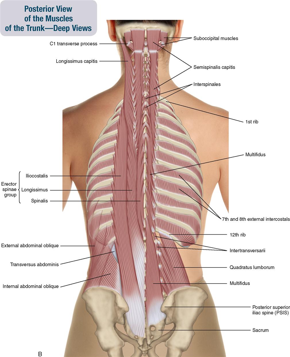
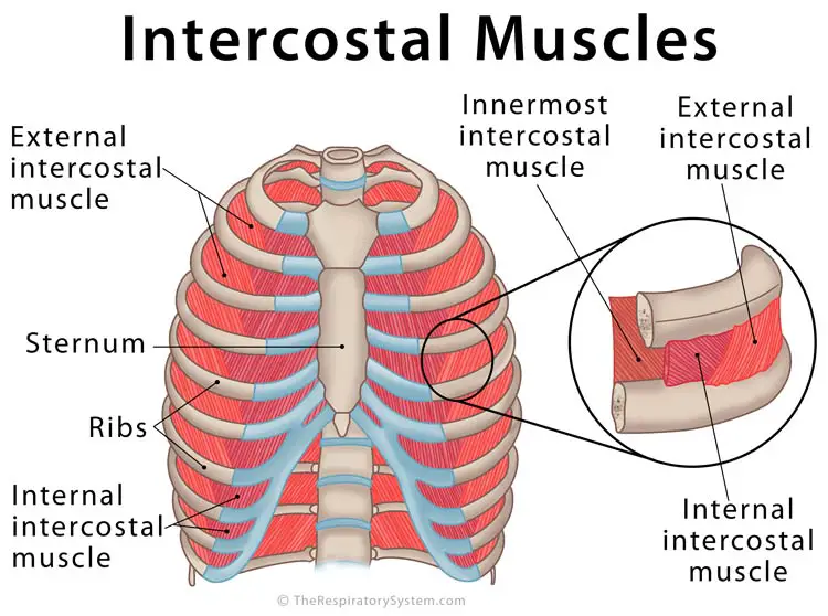
Intercostal Muscles Definition Location Anatomy Functions from www.therespiratorysystem.com The two muscles which comprise the intermediate muscle group are the serratus posterior inferior, and the serratus posterior superior. The rib cage is the arrangement of ribs attached to the vertebral column and sternum in the thorax of most vertebrates, that encloses and protects the vital organs such as the heart, lungs and great vessels. Muscles that helpful in expanding the thoracic cavity are called the inspiratory muscles because they help in inhalation, while those that compress the thoracic cavity are called expiratory. Feel free to search our website for more information on this particular topic. These muscles may be located anteriorly, posteriorly, and/or laterally. The muscles on your ribcage you are referring to are called the serratus anterior it is a muscle that originates on the surface of the 1st to 8th ribs at the side of the chest and inserts along the entire anterior length of the medial border of th. This post is about rib cage. Perform dumbbell pullovers to work the muscles along your rib cage.
Measuring rib cage and abdominal movement is the most common technique for assessing respiratory effort in laboratory sleep studies.
Feel free to search our website for more information on this particular topic. On the dorsal side there is a neural spine. They are attached to the femur (thighbone), tibia (shinbone), and fibula (calf bone) by fibrous tissues called ligaments. When you exhale, the rib cage moves down again, squeezing the air. The thoracic cage makes up the skeleton for the thoracic wall, and provides the attachments needed for the muscles of the neck, thorax. You'll need a bench and one dumbbell to do this exercise. It is formed by the vertebral column, ribs, and sternum and encloses the heart and lungs. The two muscles which comprise the intermediate muscle group are the serratus posterior inferior, and the serratus posterior superior. Rib cage diagram with organs. Whenever you bend sideways or twist your body at the hips, these muscles get called into play. Your ribs form a protective cage that encloses many of your delicate internal organs, such as your heart and lungs. The primary responsibilities of the ribcage involve protecting the thoracic visceral organs, enclosing the thoracic visceral organs, and is included in the general mechanics of the process of breathing. The rib cage has three important functions:
The rib cage has three important functions: It encloses and protects the heart and lungs. The following general rules regarding actions can be. Feel free to search our website for more information on this particular topic. Human anatomy for muscle, reproductive, and skeleton.
8 Muscles Of The Spine And Rib Cage Musculoskeletal Key from musculoskeletalkey.com When you exhale, the rib cage moves down again, squeezing the air. Whenever you bend sideways or twist your body at the hips, these muscles get called into play. Muscles that helpful in expanding the thoracic cavity are called the inspiratory muscles because they help in inhalation, while those that compress the thoracic cavity are called expiratory. Your ribs form a protective cage that encloses many of your delicate internal organs, such as your heart and lungs. If you were to develop well defined rib cage muscles, they would give off the appearance of fingers on your sides. Rib 2 is thinner and longer than rib 1 and has two articular facets on the head as normal. The other attachment of these muscles is usually considered to be either superior or inferior to the rib attachment. What you need to know.
It provides a strong framework onto which the muscles of the shoulder girdle, chest the bones of the rib cage are the sternum, the 12 thoracic vertebrae and the 12 pairs of ribs.
What you need to know. Further, there are two superior and two inferior processes meant for articulation with the neighbouring vertebra. Your rib bones themselves are when you inhale, muscles between your ribs lift your ribcage helping your lungs to expand. Rib cage diagram with organs. The thoracic cage is part of the axial skeleton (also known as the rib cage), and consists of 24 ribs, the sternum, costal cartilage, and the 12 thoracic vertebrae. The primary responsibilities of the ribcage involve protecting the thoracic visceral organs, enclosing the thoracic visceral organs, and is included in the general mechanics of the process of breathing. Perform dumbbell pullovers to work the muscles along your rib cage. The function of the rib cage is to filter the blood it receives, processing the blood. There are twelve (12) pairs of ribs and all articulate posteriorly with the thoracic vertebrae. Whenever you bend sideways or twist your body at the hips, these muscles get called into play. Muscles that helpful in expanding the thoracic cavity are called the inspiratory muscles because they help in inhalation, while those that compress the thoracic cavity are called expiratory. The ribs are a set of twelve paired bones which form the protective 'cage' of the thorax. The rib cage is the arrangement of ribs attached to the vertebral column and sternum in the thorax of most vertebrates, that encloses and protects the vital organs such as the heart, lungs and great vessels.
Your ribs form a protective cage that encloses many of your delicate internal organs, such as your heart and lungs rib cage muscles. The ribs are a set of twelve paired bones which form the protective 'cage' of the thorax.